Glaucoma is an optic nerve disease usually due to damage from high eye pressure. Sometimes when the pressure inside the eye goes up, it causes symptoms such as eye redness, eye pain, headache, and intermittent blurred vision. In other cases, there are no symptoms other than gradual loss of peripheral vision that may not be noticeable until late in the disease.
Undetected Glaucoma can lead to tunnel vision and irreversible blindness in one or both eyes. Early detection is key in prevention of vision loss in Glaucoma. At Peregrine Eye & Laser Institute (PELI), we have well-trained ophthalmologists who screen for Glaucoma using the most advanced equipment. We also offer various laser and surgical procedures in the management of Glaucoma when Glaucoma Surgery is required.

Addressing Common Glaucoma Questions
Glaucoma is damage to the optic nerve most commonly due to high eye pressure. Glaucoma causes loss of peripheral vision which if not detected and treated can eventually lead to tunnel vision and irreversible blindness.
Glaucoma can develop at any age but it is more common in persons over the age of 50. It is a disease that runs in families. Glaucoma tends to develop in individuals with high eye pressure; the normal eye pressure is from 10 to 21 mmHg.
There are two forms of Glaucoma. The first form is primary angle closure Glaucoma wherein the drainage of fluid inside the eye is blocked causing rise in eye pressure. This form of Glaucoma is more common among Asians because our eyes are smaller and the structures inside the eye are more crowded together. The second type of Glaucoma is called primary open angle Glaucoma in which although the fluid inside the eye has access to the drain, it still could not get out of the eye due to an obstruction in the deeper structures. Both types of Glaucoma result to high eye pressure. But there is another subgroup of Glaucoma under the primary open angle Glaucoma called low-tension Glaucoma. In low-tension Glaucoma, optic nerve damage and loss of peripheral vision occur in an eye wherein the eye pressure is normal.
Glaucoma causes loss of peripheral vision that may lead to tunnel vision and blindness in one or both eyes. Primary angle closure Glaucoma can cause eye redness, eye pain, headache and blurred vision that come and go. The symptoms occur when eye pressure goes up due to intermittent blockage of the drainage. In primary open angle Glaucoma, patients may not have any symptoms until late in the disease.
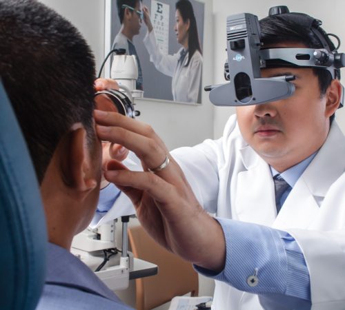
Glaucoma is diagnosed through a battery of eye tests performed by an ophthalmologist. Aside from visual acuity measurement, the peripheral field is tested using an automated perimetry machine. This is a dome-shaped machine that has a target in the center. The patient sits in front of the machine and is given a clicker. The patient is instructed to press on the clicker each time he/she sees a light flashing on the dome. A computer automatically calculates and maps the patient’s visual field. Eye pressure measurement is essential in Glaucoma screening. Eye pressure can be measured by various ophthalmic instruments. The most common is the Goldmann applanation tonometer which is mounted on a slit-lamp. Prior to eye pressure measurement, a drop of numbing agent is instilled in both eyes. This is followed by a yellow dye which will appear bright green under the blue light of the tonometer. Eye pressure measurement is not painful and takes only a few seconds to perform. Gonioscopy typically follows eye pressure measurement. In gonioscopy, a lens is placed on the patient’s eye. This lens allows the ophthalmologist to view the drainage structures of the eye. Last but not the least, the optic nerve head or optic disc is examined. The eyes may be dilated by instilling more drops before the optic discs can be examined. The ophthalmologist may use a lens to get a magnified view of the optic disc. An optical coherence tomography (OCT) scan of the optic disc is often requested during Glaucoma screening. This test determines the nerve thickness and the morphology of the optic discs. Only when all these tests are performed can an ophthalmologist diagnose with certainty if a patient has Glaucoma or not.
The goals of Glaucoma therapy are to stop optic nerve damage and halt vision loss. These are done by adequately lowering eye pressure. Once diagnosed with Glaucoma, it entails life-long, regular eye care by an ophthalmologist / Glaucoma specialist.